组会讲课人员:贺赟
Selection of Fluorescent, Bioluminescent, and Radioactive Tracers to Accurately Reflect Extracellular Vesicle Biodistributionin Vivo
准确反映细胞外囊泡体内生物分布的荧光、生物发光和放射性示踪剂的选择
主讲人:贺赟
ACS Nano|2021 Feb 23|15(2)|3212-3227 Doi: 10.1021/acsnano.0c09873
Abstract:
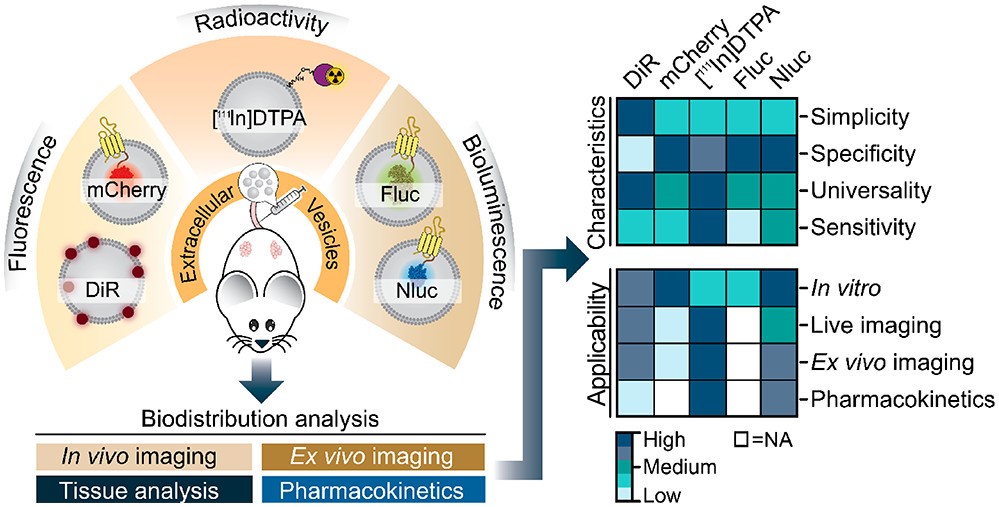
The ability to track extracellular vesicles (EVs)in vivowithout influencing their biodistribution is a key requirement for their successful development as drug delivery vehicles and therapeutic agents. Here, we evaluated the effect offive different optical and nuclear tracers on thein vivo biodistribution of EVs. Expi293F EVs were labeled using either a noncovalentfluorescent dye DiR, or covalent modification with111indium-DTPA, or bioengineered withfluorescent (mCherry) or bioluminescent (Firefly and NanoLuc luciferase) proteins fused to the EV marker, CD63. To focus specifically on the effect of the tracer, we compared EVs derived from the same cell source and administered systemically by the same route and at equal dose into tumor-bearing BALB/c mice.111Indium and DiR were the most sensitive tracers forin vivoimaging of EVs, providing the most accurate quantification of vesicle biodistribution byex vivoimaging of organs and analysis of tissue lysates. Specifically, NanoLuc fused to CD63 altered EV distribution, resulting in high accumulation in the lungs, demonstrating that genetic modification of EVs for tracking purposes may compromise their physiological biodistribution. Blood kinetic analysis revealed that EVs are rapidly cleared from the circulation with a half-life below 10 min. Our study demonstrates that radioactivity is the most accurate EV tracking approach for a complete quantitative biodistribution study including pharmacokinetic profiling. In conclusion, we provide a comprehensive comparison offluorescent, bioluminescent, and radioactivity approaches, including dual labeling of EVs, to enable accurate spatiotemporal resolution of EV trafficking in mice, an essential step in developing EV therapeutics.
.
摘要:
在不影响细胞外囊泡生物分布的情况下,能够在活体内追踪细胞外囊泡是其作为药物载体和治疗剂成功开发的关键。在这里,我们评估了5种不同的光学和核示踪剂对EVS体内生物分布的影响。Expi293F EV用非共价荧光染料DIR标记,或用111铟-DTPA共价修饰,或用荧光(MCherry)或生物发光蛋白(Firefly和NanoLuc荧光素酶)融合到EV标记CD63上进行生物工程标记。为了特别关注示踪剂的效果,我们比较了来自相同细胞的EV,并以相同的途径和相同的剂量系统地给药到荷瘤的BALB/c小鼠中。111铟和DIR是EV活体成像的最敏感的示踪剂,通过器官的体外成像和组织裂解物的分析提供了最准确的囊泡生物分布的量化。具体地说,NanoLuc与CD63融合改变了EV的分布,导致EV在肺部的高蓄积,表明出于跟踪目的对EV的基因改造可能会损害它们的生理生物分布。血液动力学分析显示,EV迅速从循环中清除,半衰期低于10分钟。我们的研究表明,放射性是包括药代动力学研究在内的完整定量生物分布研究中最准确的EV跟踪方法。总之,我们对荧光、生物发光和放射性方法进行了全面的比较,包括EV的双重标记,以实现EV在小鼠体内运输的准确时空分辨率,这是开发EV治疗的关键步骤。